Jason Huang

The Achilles tendon is one of the strongest and toughest tendons in the human body. Yet, strangely, many Achilles tendon ruptures are not caused by an impact, but simply by a single stride, push-off, jump, or tiptoeing motion. Kevin Durant suffered an Achilles tendon rupture at age 30, and Kobe Bryant faced the same injury at age 34. People in their 30s are clearly still young, so why does the Achilles tendon suddenly rupture?
Does the Achilles tendon degenerate by age 30?!
Studies analyzing spontaneously ruptured tendons have found that the vast majority actually had varying degrees of degenerative tissue changes prior to the rupture (Kannus & Józsa, 1991). In other words, that final step might just be the straw that broke the camel's back.
The 30s is a particularly noteworthy stage. The muscles are still very strong, and speed and explosive power are still excellent; however, the Achilles tendon has already endured ten to twenty years of running, jumping, sudden stops, and pushing off. The tendon bundles within the Achilles tendon need to slide against each other, stretch, and recoil to store and release energy, and these tissue characteristics gradually change with age and long-term loading (Thorpe et al., 2015). Therefore, a situation may arise where the muscles are still strong, but the tissue responsible for transmitting force has already begun to degenerate. But why does the tendon degenerate so early?
The problem may not lie solely with the Achilles tendon
The human body is not just one muscle connected to one tendon. In fact, the soft tissue on the back of the lower leg is a continuous system: plantar fascia → calcaneus → Achilles tendon → soleus → gastrocnemius → posterior knee. A 2024 ultrasound study found that patients with Achilles tendinopathy not only exhibited changes in the Achilles tendon and paratenon tissue, but the deep fascia between the gastrocnemius and soleus muscles was also significantly thickened (Ryskalin et al., 2024). This indicates that when problems arise in the Achilles tendon, we perhaps shouldn't look at the Achilles tendon in isolation.
My Hypothesis: The "Soft Tissue Space" Might Be the First to Change
This is also one of the research directions of the Soft Tissue Deformation Theory that I proposed. When forces from running, jumping, pushing off, and sudden stops act repeatedly over many years, combined with static sitting and exerting force in a shortened state, the soft tissues on the back of the lower leg may not just "get tight," but gradually undergo changes in their alignment and spatial distribution.
Anyone who has taken my classes knows that I pay special attention to the fascia around joints, bone ends, and bony clefts. For the lower leg, this means the ankle joint, calcaneus, and the popliteal space behind the knee. My hypothesis is that if the soft tissues on the back of the lower leg undergo long-term deformation toward the popliteal space and ankle joint clefts, the upper fascial tissues are gradually compressed, while the lower fascia may be continuously stretched, further altering the original tension and sliding relationship among the gastrocnemius, soleus, and Achilles tendon (Figure 1). This may also affect local pressure, interstitial fluid exchange, microcirculation, and the cellular metabolic environment. Furthermore, the blood supply to the Achilles tendon itself is not abundant, and relatively hypovascular zones are also thought to be associated with tendon degeneration and repair capacity (Ahmed et al., 1998). Therefore, tightness represents a lack of space, cramped space leads to poorer circulation, and day after day, a rupture occurring in the 30s is actually a reasonable consequence.
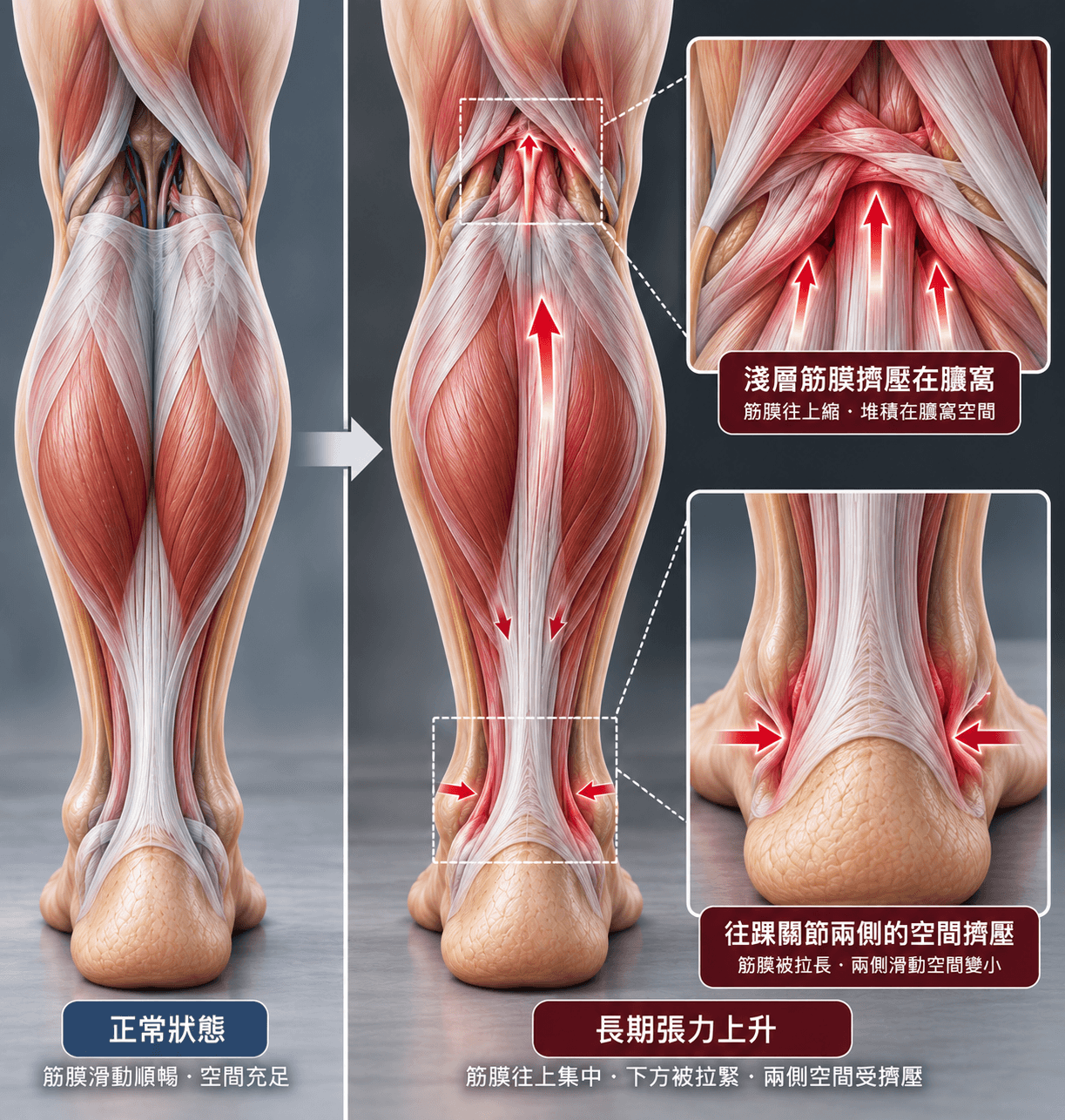
Figure 1: Schematic diagram of the mechanism where fascial tissues on the back of the lower leg deform due to exercise and sedentary lifestyles, leading to tightness.
Therefore, I propose a possible mechanistic pathway worthy of further study: long-term soft tissue deformation - altered fascial alignment and sliding - altered local tension and pressure - potential impact on circulation and metabolism - gradual accumulation of micro-damage - decreased tolerance of the Achilles tendon - final push-off - rupture (Figure 2).
Current research has found an association between Achilles tendinopathy and abnormalities in the upstream intermuscular fascia, but the hypothesis that "fascial deformation causes circulatory changes, further leading to Achilles tendon rupture" still requires scientific validation. (Ryskalin et al., 2024)
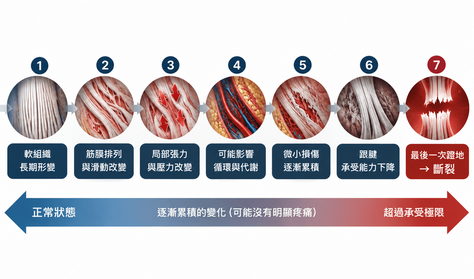
Figure 2: Flowchart of the hypothetical physiological mechanism of Achilles tendon rupture.
How do you know if your Achilles tendon might need attention?
Don't wait until you feel Achilles tendon pain to start observing. You can pay attention to several things in your daily routine:
1. Observe the lower legs: Check if the appearance and muscle belly position of the left and right gastrocnemius muscles, as well as the soft tissue distribution behind the knees, are significantly different.
2. Observe the ankle joints: Are the dorsiflexion angles of the left and right feet different? Does one heel lift particularly easily when squatting?
3. Perform single-leg heel raises: Compare whether the height of the heel raises is consistent on both sides. The lower side may require some maintenance.
4. Palpate the Achilles tendon: Is there thickening, tenderness, swelling, or a difference in thickness between the left and right sides?
5. Look at the recovery rate: Used to recover the day after exercise, but now frequently experience calf stiffness? Does Achilles tendon tightness start to appear with the first step in the morning?
These phenomena do not mean "the Achilles tendon is about to rupture"; they simply indicate that the soft tissues have already begun to change. However, if pain, swelling, or decreased strength persist, you should seek professional evaluation.
Maintain your Achilles tendon, but don't only maintain the Achilles tendon
In professional horse racing, it's not just about resting after a run. The training team, usually consisting of equine veterinarians, trainers, rehabilitation/physical therapy-related personnel, and horse caregivers, collectively observes the state of the limbs' tendons. They perform manual palpation after training daily to check for localized heat, swelling, pain, or differences between left and right sides in the tendons. When further confirmation is needed, veterinarians use musculoskeletal ultrasound to observe tendon thickness, fiber alignment, and the presence of lesions. Recovery management may include manual or tool-assisted massage, ice application, or cold therapy equipment, with training load and recovery time adjusted based on the tendon's condition. But what about us as humans?
Many people only stretch when they feel tight, treat it only when they feel pain, or even start prioritizing the Achilles tendon only after it ruptures. Athletes all know the importance of stretching, but if stretching alone were enough to solve all problems, why do professional athletes who train regularly and stretch daily still experience Achilles tendon ruptures? I believe what truly needs to be managed might not just be "flexibility," but also the tension, sliding, space, and load of the entire plantar fascia-Achilles tendon-calf-posterior knee soft tissue system.
Just as we regularly manage blood pressure, blood sugar, and blood lipids, soft tissue tension also needs regular observation and restoration. When long-term training increases fascial tension, reduces tissue sliding, and even gradually causes deformation in alignment and space, simply "elongating" the muscle may not be enough.
Being in your 30s is still very young. But body maintenance cannot wait until you are old to begin.
Disclaimer
The content of this article is for health education and knowledge sharing purposes regarding exercise, fascia, and body maintenance only, and is not intended as a disease diagnosis, treatment, or individual medical advice. Some views in the text regarding soft tissue deformation, fascial tension, and tissue space are theories and research hypotheses proposed by the author, which still require more scientific research for validation. If you experience persistent pain, swelling, limited mobility, decreased muscle strength, or suspect an Achilles tendon injury, please seek evaluation and treatment from a qualified physician or relevant medical professional.


